At Long Island Neurology Consultants, we perform a vast range of neurological testing at our Lynbrook office.
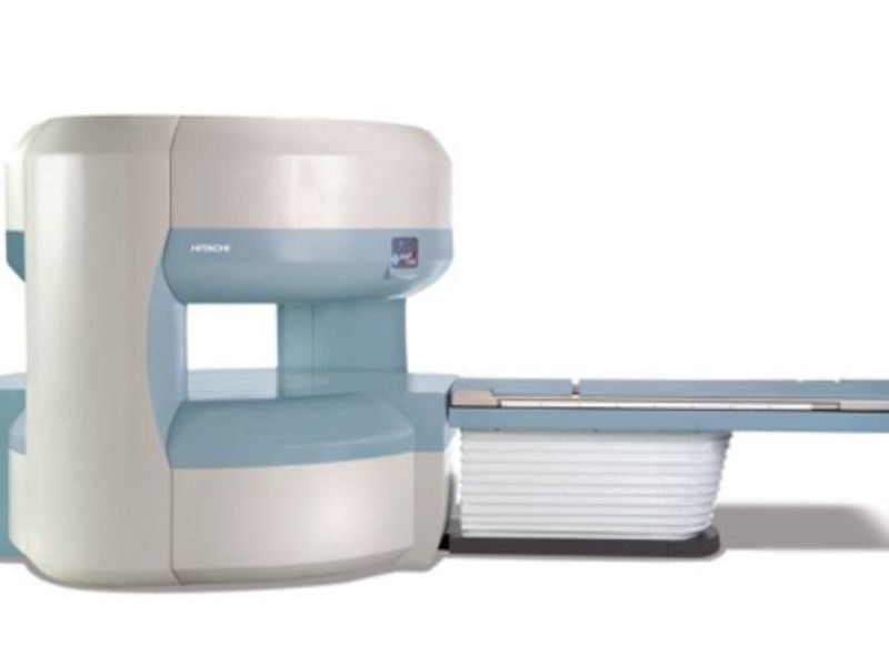
Magnetic Resonance Imaging (MRI)
MRI is a testing modality that uses pulses of energy and magnetic fields to image the body. MRI is highly sensitive and can provide more specific information regarding the nervous system than other testing modalities such as ultrasound, X-ray, or computed tomography (CT).
Please note, MRI does NOT expose a patient to radiation. In neurology, we use MRI most commonly to look at detailed anatomy of the brain and spinal cord.
For more information on MRI and how to prepare for it,click here.
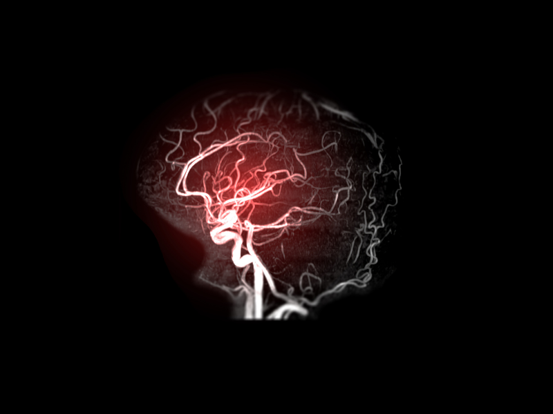
Magnetic Resonance Angiography (MRA) / Magnetic Resonance Venography (MRV)
MRA is a testing modality that is utilized to image arteries. From a patient standpoint, it is very similar to having an MRI. MRA does NOT expose a patient to radiation. In most cases, contrast is not needed. MRA can provide more specific information about the vascular system than other testing modalities including computer tomography (CT), X-ray, and ultrasound.
In neurology, we use MRA most commonly to look at arteries of the brain and neck to assess for abnormalities such as aneurysms, malformations, and narrowing.
Magnetic Resonance Venography (MRV)
This study is similar to MRA, but focuses on the veins.
For more information on MRA and how to prepare for it,click here.
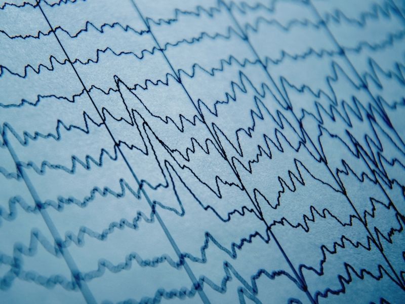
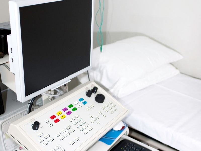
Electroencephalogram (EEG)
An Electroencephalogram (EEG) records the electrical activity of the brain. Our certified technologist will measure the patient’s head and put the electrodes on their scalp with a paste-like substance. During the test, the patient lies on a table (or may remain in a chair) in a dark room, remaining still and relaxed. Sleep is encouraged during the procedure. Often, a strobe light is used as well to record the brain’s response. An EEG usually takes between 30 to 45 minutes.
For more information on EEG and how to prepare for it,click here.
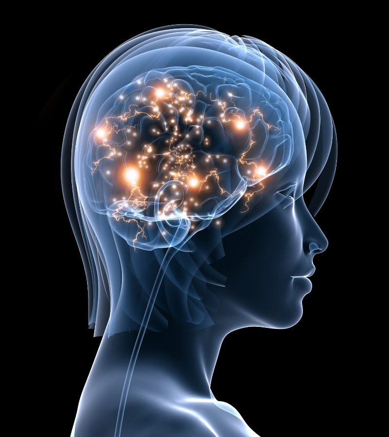
Evoked Potential (EP)
Our office performs four different Evoked Potential (EP) examinations. Testing is performed by a trained technician. Each test may take about 20 minutes. Often, several EP’s are scheduled during a session.
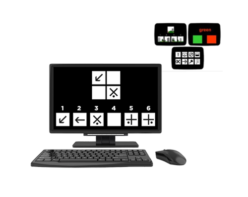
Visual Evoked Potential (VEP or VER for visual evoked response):
This test evaluates the visual portion of the nervous system from the eyes to the occipital (visual) portion of the brain. Electrodes are pasted to the scalp, and the patient stares at a checkerboard pattern on a video screen. Each eye is tested separately.
Brainstem Auditory Evoked Potential (BAEP or BAER for brainstem auditory evoked response):
This test evaluates the auditory portion of the nervous system from the ears through the brainstem and the auditory portion of the brain. Electrodes are pasted to the scalp, and the patient wears headphones which deliver a series of clicks and tones to each ear separately. This is NOT the same as a hearing test (or audiogram), and it provides different information.
Somatosensory Evoked Potentials (SSEP):
There are two different forms of the examination to assess either the upper extremities or the lower extremities. Both tests assess pathways from the limb through the spinal cord and to the brain. Electrodes are pasted to the skin and scalp. While the patient remains in a relaxed position, a small electrical current is applied to the skin, creating a tingling sensation.
For more information on this exam and how to prepare for it,click here.
NeuroTrax
NeuroTrax is a cloud-based computer examination used to assess cognition, including memory. The test does NOT require specific computer skills. It is administered by our staff who will assist you throughout the testing session. The testing sessions are between 40 to 75 minutes in length. There may be times when testing sessions run longer depending on the patient’s condition.
For More information on NeuroTrax and how to prepare, click here.
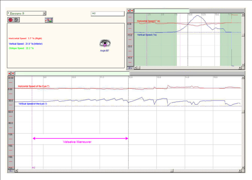
Video Electronystagmography (VNG)
Video electronystagmography (VNG or VENG) is a test for assessing dizziness. During the exam, the patient will sit in a dark room and wear infrared googles which have a camera to record eye movements known as nystagmus.
The procedure averages between 45 to 60 minutes.
For more information on this exam and how to prepare for it,click here.
Electromyography/nerve conduction studies (more typically referred to as just EMG) is a form of electrodiagnostic testing that is used to study nerve and muscle function. Our studies are performed by a physician with specialized training for this procedure.
The study consists of two parts, both of which are performed by the doctor. In the first part (nerve conduction study), small discs known as electrodes are taped to the skin. The nerve is then electrically stimulated to evaluate its strength and speed. In the second part (electromyography), a small wire electrode is inserted into several muscles to listen to muscle activity both at rest and while you move the muscle.
The procedure averages between 30 to 90 minutes depending on the number of extremities being tested. The physician will explain each part of the examination as it is being performed. If you have any concerns during the procedure, please alert the physician who can modify the exam as needed.
For more information on EMG and how to prepare for it,click here.
Our neurovascular laboratory is certified by the IAC (Intersocietal Accreditation Commission). The IAC is a nationally recognized accrediting organization to advance appropriate utilization, standardization, and quality of diagnostic imaging. All our sonographers are nationally registered vascular technologists (RVT) with credentials in vascular imaging which are updated annually.
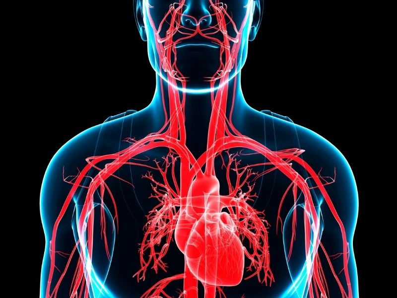
Carotid Doppler
Carotid Doppler(Carotid Ultrasound) is a diagnostic procedure used to examine the carotid arteries for increased stroke risk. Ultrasound is widely used and captures images in real-time. This non-invasive test is performed by our registered vascular technologist. The patient sits in a reclining chair while the technologist applies a gel to a flat probe moving up and down the neck, taking images of the carotid arteries along the way. The procedure averages about 30 minutes.
For more information on this exam and how to prepare for it,click here.
Transcranial Doppler, Embolic Screening/VasoReactivity Study
Transcranial Doppler with Embolic Screening/Vasoreactivity Study
Similar to carotid doppler examination, this non-invasive diagnostic procedure examines the arteries of the head for increased stroke risk. The registered vascular technologist will apply a gel to a flat probe placing it on several areas of the head including the eyelids, temples, and back of the neck. It is often performed at the same time as the carotid doppler and both procedures typically take 45 minutes combined.
For more information on this exam and how to prepare for it,click here.